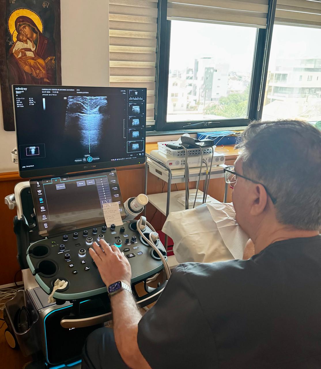
Lung ultrasound (LUS) has become one of the most valuable bedside imaging techniques in modern cardiology, emergency medicine, internal medicine, and critical care. It is rapid, radiation-free, repeatable, and significantly more sensitive than chest radiography for many pulmonary conditions.For a cardiologist, lung ultrasound is now considered an essential part of comprehensive cardiovascular assessment because it provides real-time information regarding pulmonary congestion, pleural disease, and many lung pathologies.
What Can Lung Ultrasound Diagnose?
1. Pulmonary Congestion (Heart Failure)
This is probably the most important application in cardiology.
Findings:
- Multiple bilateral B-lines
- Diffuse interstitial syndrome
- Pleural effusions
- Reduced lung sliding only if another pathology coexists
Clinical value:
- Detects pulmonary edema before symptoms become severe.
- Estimates congestion much better than chest X-ray.
- Helps titrate diuretics.
- Predicts hospitalization and prognosis.
- Useful during follow-up after treatment.
2. Pleural Effusion
Findings:
- Anechoic fluid above the diaphragm
- Floating lung ("jellyfish sign")
- Compressive atelectasis
Can estimate:
- Volume
- Location
- Complexity
- Septations
3. Pneumonia
Findings:
- Consolidation
- Dynamic air bronchograms
- Irregular pleural line
- Focal B-lines
Sensitivity is approximately 90–95%, considerably higher than chest radiography in many patients.
4. Pneumothorax
Characteristic signs:
- Absent lung sliding
- Absent B-lines
- Lung point (highly specific)
- Barcode/Stratosphere sign on M-mode
5. Interstitial Lung Disease
Findings:
- Numerous B-lines
- Thickened pleural line
- Pleural irregularity
- Subpleural