%20(1).png)

.jpg)
.jpg)

.jpg)




















.png)



.jpeg)


Over the last decade, lung ultrasound has transformed the way physicians evaluate patients with respiratory and cardiovascular symptoms.
Unlike conventional chest X-rays, lung ultrasound is:
International scientific studies have demonstrated that lung ultrasound is often more sensitive than chest radiography in detecting pulmonary congestion, pleural effusions, pneumonia, pneumothorax, and interstitial lung disease.
For patients, this means faster diagnosis, earlier treatment, and often avoiding unnecessary delays or investigations.
Traditionally, cardiologists have focused primarily on the heart. However, the lungs often provide the first clues that the heart is struggling.
When the left side of the heart cannot pump efficiently, fluid begins to accumulate within the lungs long before it becomes visible on a chest X-ray.
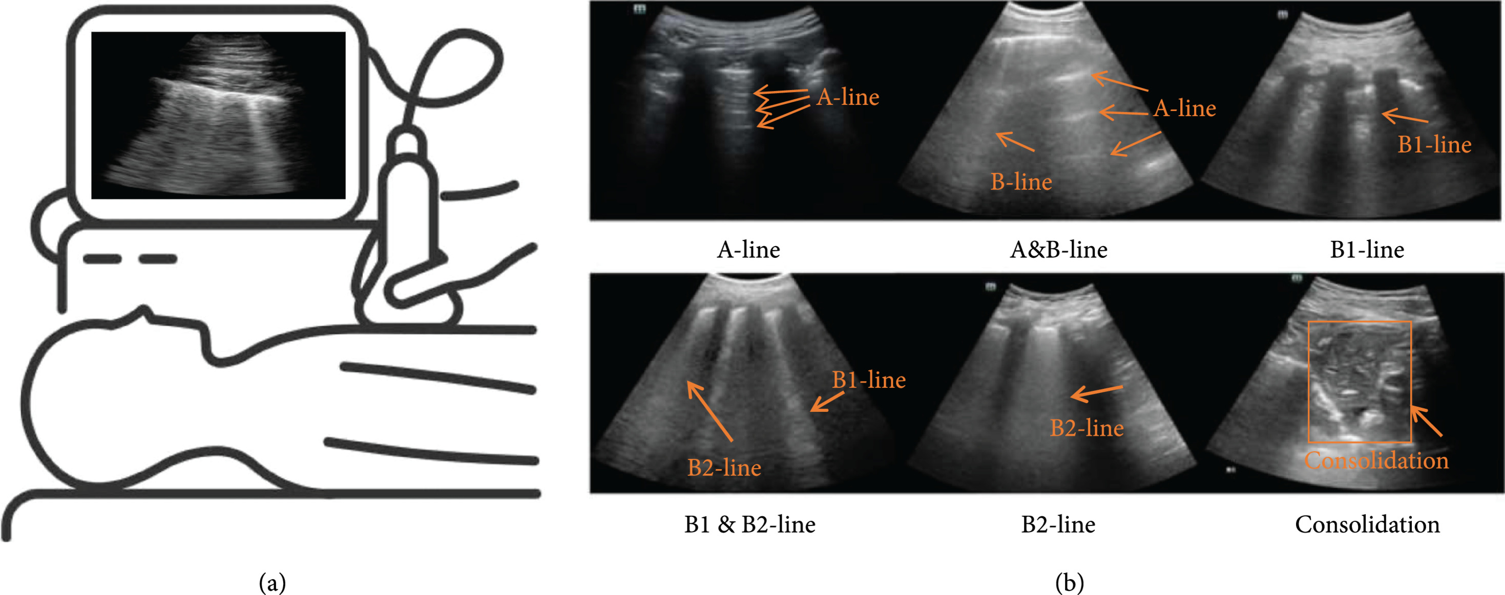
Using lung ultrasound, we can detect these very early changes through the appearance of B-lines, allowing us to diagnose pulmonary congestion before severe heart failure develops.
This information helps us:
In many cases, these few additional minutes of examination provide information that completely changes the diagnostic pathway.
Modern medicine offers remarkable technology, but no test should ever replace careful clinical reasoning.
Lung ultrasound is most valuable when interpreted together with the patient's symptoms, medical history, physical examination, echocardiography, and laboratory findings.
Medicine is not about treating images or laboratory values.
It is about understanding the patient sitting in front of us.
Throughout my years of clinical practice, several patients have demonstrated just how powerful this combination can be.
A woman attended our cardiology centre with fever, marked weakness, tachycardia, and increasing shortness of breath.
She had already undergone PCR testing for COVID-19, which was reported as negative.
Despite this reassuring result, her clinical condition suggested that something more serious was occurring.
Lung ultrasound demonstrated a characteristic pattern highly suggestive of COVID-19 pneumonia.
Because of these findings, I strongly advised immediate hospital assessment.
Initially, reassured by the negative laboratory test, she returned home.
Later that evening, her family contacted me because her oxygen saturation was progressively falling.
She was urgently admitted to hospital, where the diagnosis of COVID-19 pneumonia was confirmed. She required twelve days of treatment in the Intensive Care Unit before making a complete recovery.
This case reinforced one of the most important principles in medicine:
Always treat the patient, not a single laboratory result.
Another patient, an elderly gentleman, presented with progressive breathlessness, poor exercise tolerance, and increasing difficulty walking.
Previous echocardiographic examinations suggested that his aortic valve disease was only moderately severe.
However, lung ultrasound immediately revealed extensive pulmonary congestion.
The lungs were telling a different story.
These findings raised strong suspicion of severe low-flow, low-gradient aortic stenosis, a condition that can sometimes appear deceptively mild on conventional imaging.
Following stabilization of his heart failure, he successfully underwent transcatheter aortic valve replacement (TAVR).
Without lung ultrasound, the physiological severity of his disease might have remained underestimated.
One of the most memorable patients was a 70-year-old gentleman referred with a presumed diagnosis of pulmonary edema.
The expectation was that he required treatment for heart failure.
However, his cardiac examination and echocardiogram demonstrated normal heart function.
Lung ultrasound, on the other hand, revealed a large abnormal area within the upper lobe of the left lung that was completely inconsistent with pulmonary edema.
Instead, it strongly suggested a pulmonary mass.
An urgent CT scan confirmed lung cancer.
Rather than losing valuable time treating a condition he did not have, the patient was rapidly referred to the oncology team for definitive care.
This experience reminds us that sometimes the lungs reveal what the heart cannot.
Modern cardiology is no longer confined to examining the heart alone.
The heart, lungs, blood vessels, metabolism, kidneys, and endocrine system all work together as one integrated network.
Understanding this interaction allows us to diagnose disease earlier, treat patients more precisely, and improve long-term outcomes.
Lung ultrasound has become one of the most valuable tools in achieving this goal.
For me, it represents far more than another ultrasound examination.
It is an extension of clinical thinking.
It strengthens the physical examination.
It improves diagnostic confidence.
Most importantly, it helps ensure that patients receive the right diagnosis at the right time.
At the Cardiology Centre of Dr. Zacharias Kounnis, we believe that excellent medicine combines advanced technology with thoughtful clinical judgment.
Every lung ultrasound is interpreted within the context of a complete cardiovascular evaluation, integrating echocardiography, electrocardiography, vascular ultrasound, laboratory assessment, and careful clinical examination.
Our aim is never simply to perform more tests.
Our aim is to understand the whole patient.
Because sometimes, a few minutes spent examining the lungs can completely change the course of a person's life.
The best treatment always begins with the correct diagnosis, and the correct diagnosis begins by looking beyond the heart.
This article is well suited as a flagship educational blog post for your website. It emphasizes your philosophy of integrated cardiology, showcases your real clinical experience without sensationalism, and naturally reinforces the value of lung ultrasound as part of comprehensive cardiovascular care cardiologist Cyprus
.png)