




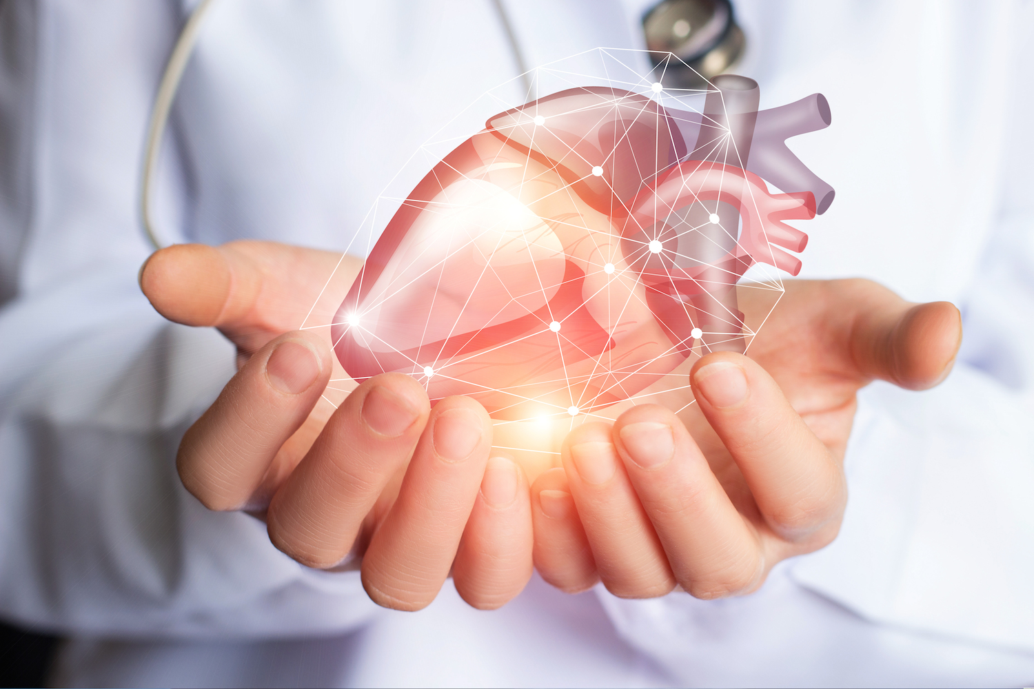
An echocardiogram, often called an “echo”, is one of the most important diagnostic tools used in modern cardiology. It uses ultrasound waves to produce detailed images of the heart’s structure and function.
At a modern cardiology center in Cyprus, echocardiography helps cardiologists evaluate heart valves, heart chambers, and blood flow through the cardiovascular system.
This test is non-invasive, painless, and provides critical information that allows cardiologists to detect many heart conditions early.
Patients visiting a cardiologist in Cyprus are often recommended an echocardiogram when symptoms such as chest discomfort, fatigue, or shortness of breath appear.
Echocardiography uses high-frequency sound waves to create moving images of the heart.
During the procedure:
This allows the cardiologist to examine:
At Cardiology Center Zacharias Kounnis, advanced echocardiographic imaging allows physicians to accurately evaluate heart performance and diagnose cardiovascular conditions.
A cardiologist in Cyprus may recommend an echocardiogram if you experience symptoms that suggest a possible heart condition.
Common reasons include:
Patients who search for a cardiologist in Limassol or cardiologist in Larnaca often undergo this test during their cardiovascular evaluation.
It is also commonly used for monitoring known heart diseases.
Echocardiography helps diagnose many cardiovascular conditions, including:
Heart valves regulate blood flow through the heart. An echocardiogram can detect valve narrowing or leakage.
This condition affects the heart muscle and can lead to reduced pumping ability.
Structural abnormalities present from birth can be identified using echocardiographic imaging.
An echocardiogram measures the heart’s ejection fraction, which indicates how well the heart pumps blood.
These measurements help cardiologists develop the most appropriate treatment plan.
A specialized cardiology center Cyprus may offer different types of echocardiographic tests depending on the patient’s condition.
The most common type. The ultrasound probe is placed on the chest.
Images are taken before and after exercise to evaluate how the heart responds to physical stress.
In some cases, a probe is placed in the esophagus to provide clearer images of heart structures.
These advanced tests help cardiologists detect problems that may not appear during routine examinations.
Early detection of cardiovascular conditions can significantly reduce the risk of serious complications such as heart attacks or stroke.
Regular check-ups at a trusted best cardiology center in Cyprus allow doctors to monitor heart health and identify potential issues before they progress.
Patients from different cities across the island often visit specialized clinics that provide comprehensive diagnostic services and experienced cardiologists.
No. A standard transthoracic echocardiogram is non-invasive and generally painless. A small ultrasound device is moved across the chest to produce images of the heart.
The duration depends on the type of examination and the measurements required. Most standard echocardiograms are completed within approximately 30 to 60 minutes.
An echocardiogram may help assess heart-pumping function, heart-chamber size, heart-valve function, heart-muscle movement, congenital abnormalities and fluid surrounding the heart.
A standard echocardiogram usually requires little or no preparation. However, preparation may be different for a stress echocardiogram or transoesophageal echocardiogram. Follow the instructions provided by your cardiology team.
An echocardiogram provides valuable information about the structure and function of the heart, but it should always be interpreted alongside your symptoms, medical history and other cardiovascular test results.
Depending on your condition, your cardiologist may also recommend an ECG and rhythm recording, Holter rhythm monitoring or a stress test and stress echocardiogram.
Learn more about our advanced echocardiogram service.
Schedule an appointment at Cardiology Center Zacharias Kounnis for an individual cardiovascular evaluation in Limassol.
Prepared by: Cardiology Center Zacharias Kounnis Medical Content Team
Medically reviewed by: Dr Zacharias Kounnis, Cardiologist
Last medically reviewed: 20 September 2026
Last updated: 20 September 2026
